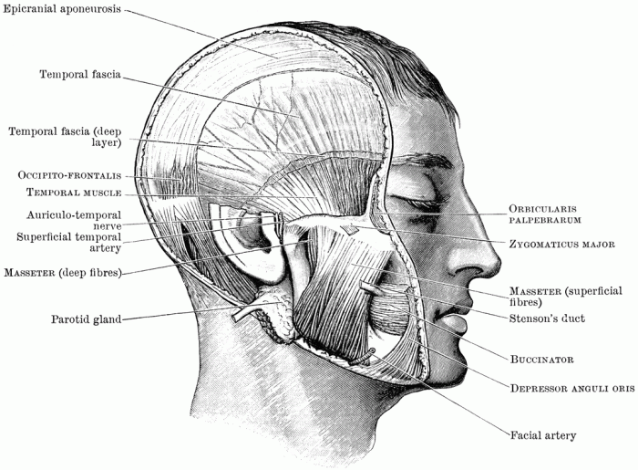
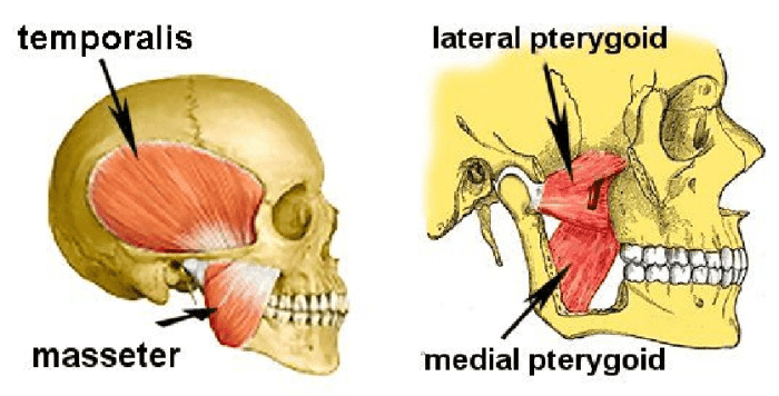
Label the muscles of mastication in the figure. This article delves into the intricacies of the muscular apparatus responsible for mastication, the crucial first step in the digestive process. We explore the location, innervation, and function of each muscle, highlighting their coordinated actions in achieving efficient chewing.
Introduction

The muscles of mastication are a group of muscles that are responsible for chewing. They are located in the head and neck and are innervated by the trigeminal nerve. Mastication is the process of breaking down food into smaller pieces so that it can be more easily swallowed and digested.
It is an important part of the digestive process and is essential for proper nutrition.
Muscles of Mastication: Label The Muscles Of Mastication In The Figure.

The primary muscles involved in mastication are the masseter, temporalis, medial pterygoid, and lateral pterygoid muscles.
- Masseter muscle:The masseter muscle is a thick, square muscle that is located on the side of the face. It originates from the zygomatic arch and inserts on the mandible. It is innervated by the mandibular nerve.
- Temporalis muscle:The temporalis muscle is a fan-shaped muscle that is located on the side of the head. It originates from the temporal fossa and inserts on the mandible. It is innervated by the mandibular nerve.
- Medial pterygoid muscle:The medial pterygoid muscle is a small, triangular muscle that is located on the medial side of the mandible. It originates from the medial pterygoid plate and inserts on the mandible. It is innervated by the mandibular nerve.
- Lateral pterygoid muscle:The lateral pterygoid muscle is a large, triangular muscle that is located on the lateral side of the mandible. It originates from the lateral pterygoid plate and inserts on the mandible. It is innervated by the mandibular nerve.
Function of Muscles
The muscles of mastication work together to achieve efficient chewing. The masseter and temporalis muscles close the jaw, while the medial and lateral pterygoid muscles move the jaw from side to side and up and down. This allows the teeth to grind and break down the food into smaller pieces.
Clinical Relevance

An understanding of the muscles of mastication is crucial in diagnosing and treating disorders related to mastication. For example, temporomandibular joint (TMJ) disorders are a group of conditions that affect the jaw joint and can cause pain, clicking, and difficulty chewing.
These disorders can be caused by a variety of factors, including muscle imbalances, trauma, and arthritis.
Question Bank
What are the primary muscles involved in mastication?
The primary muscles involved in mastication are the masseter, temporalis, medial pterygoid, and lateral pterygoid muscles.
How do the muscles of mastication work together?
The muscles of mastication work in a coordinated fashion to achieve efficient chewing. The masseter and temporalis muscles elevate the mandible, while the medial and lateral pterygoid muscles protrude and retract the mandible, respectively.